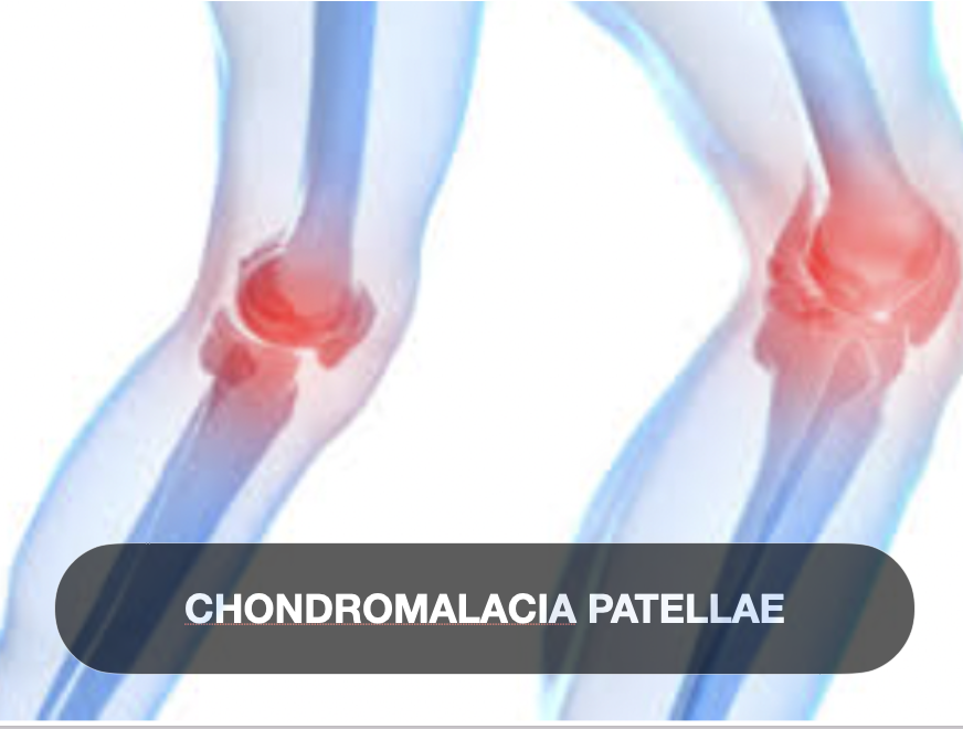
Chondromalacia Patellae
Introduction
Chondromalacia patellae is a common cause of anterior knee pain, especially among young and active individuals. It involves the softening, degeneration, and sometimes fibrillation of the articular cartilage on the undersurface of the patella (kneecap). This condition disrupts normal patellofemoral joint biomechanics, often resulting in pain and functional limitations. Understanding the pathology, underlying causes, classification systems, and clinical features is vital for physiotherapists to develop targeted rehabilitation strategies aimed at symptom relief and functional restoration.
Definition
Chondromalacia patellae refers to the degenerative changes and softening of the hyaline cartilage on the posterior (undersurface) aspect of the patella.
It is characterized histologically by cartilage matrix softening, swelling, fibrillation, and eventual cartilage thinning or erosion.
The condition is often considered part of the spectrum of patellofemoral pain syndrome (PFPS) but with a more defined cartilage pathology.
It is distinct from osteoarthritis but can progress to degenerative joint disease if untreated.
Cause
Biomechanical factors leading to abnormal patellofemoral joint stress are the primary cause:
Malalignment of the patella (e.g., lateral tracking or tilt).
Muscle imbalances, particularly weakness of the vastus medialis oblique (VMO) relative to the vastus lateralis.
Overuse or repetitive microtrauma from activities involving frequent knee flexion and extension such as running, jumping, or cycling.
Direct trauma to the patella can initiate cartilage damage.
Anatomical variations like trochlear dysplasia or patella alta (high-riding patella) contribute to uneven load distribution.
Age-related wear and cartilage degeneration can also play a role, though chondromalacia is more common in younger individuals compared to osteoarthritis.
Risk factors include overweight status, poor lower limb biomechanics, and previous knee injuries.
Classification
Chondromalacia patellae is often classified according to the Outerbridge grading system based on arthroscopic findings:
Grade I: Softening and swelling of the cartilage without surface disruption.
Grade II: Fibrillation or fragmentation affecting less than 1.5 cm².
Grade III: Fibrillation and fissuring extending more than 1.5 cm² but without full-thickness loss.
Grade IV: Exposed subchondral bone due to full-thickness cartilage loss.
This classification helps guide prognosis and treatment planning.
Signs and Symptoms
Anterior knee pain: Typically diffuse, poorly localized around or behind the patella; worsens with activities that increase patellofemoral joint load (e.g., stair climbing, squatting, prolonged sitting).
Crepitus: A grinding or cracking sensation felt during knee movement, especially flexion-extension.
Tenderness: Palpation around the patella and along the joint margins reproduces pain.
Swelling: Mild joint effusion may be present in some cases.
Reduced function: Difficulty with weight-bearing activities and a feeling of instability or weakness around the knee.
Pain during resisted knee extension and positive clinical tests like the Clarke’s sign (patellar grind test) may assist diagnosis.
Symptoms often worsen with prolonged sitting (the “theater sign”) or after intense activity.
Conclusion
Chondromalacia patellae is a prevalent and clinically significant condition characterized by degenerative cartilage changes in the patellofemoral joint, leading to anterior knee pain and functional impairment. Early recognition and a thorough understanding of its biomechanical causes and clinical presentation enable physiotherapists to design effective, individualized rehabilitation programs. Emphasizing quadriceps strengthening, patellar realignment techniques, and activity modification can improve symptoms and prevent progression to osteoarthritis.

