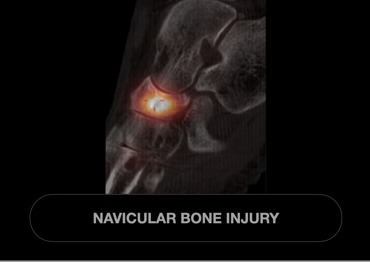
Navicular Bone Injury .
Ouch
Navicular bone injuries primarily include
Fractures - Acute navicular fractures are commonly caused by direct trauma or severe twisting injuries, frequently seen in athletes.
Classification systems for navicular fractures (e.g., Sangeorzan classification) help guide treatment, ranging from non-displaced fractures managed conservatively to displaced fractures requiring surgical intervention.
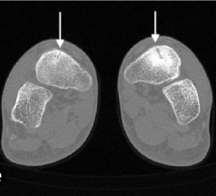
Stress fractures - often associated with high-impact activities or repetitive stress. Stress fractures of the navicular bone occur due to repetitive microtrauma, particularly in runners and dancers, presenting with midfoot pain and tenderness.
Delayed diagnosis of navicular stress fractures can lead to nonunion or avascular necrosis due to the bone’s limited blood supply.
Accessory Bone -Os Tibiale Externum is an accessory bone located near the medial side of the navicular bone in the foot. It is a congenital anatomical variant, often asymptomatic but can sometimes cause medial foot pain or discomfort due to irritation or inflammation. Diagnosis is typically confirmed through imaging, such as X-rays. Treatment ranges from conservative management with orthotics and anti-inflammatory measures to surgical excision in refractory cases. Understanding this condition is crucial for accurate diagnosis and appropriate intervention to alleviate symptoms. or
Accessory Bone - Os Naviculare is an accessory bone located near the navicular bone in the foot. It is a congenital anatomical variant present in some individuals and usually asymptomatic. However, it can sometimes cause pain or discomfort due to inflammation or injury, particularly in athletes or those experiencing repetitive stress. Diagnosis is typically confirmed through imaging, and treatment ranges from conservative management like rest and orthotics to surgical intervention in persistent cases.

Navicular Stress Fractures
Introduction
Navicular stress fractures are a significant concern within the realm of lower extremity injuries, particularly impacting athletes and individuals engaged in repetitive high-impact activities. Understanding the nature of this specific injury, its underlying causes, and the clinical manifestations is essential for timely diagnosis and effective management. This summary synthesises current medical literature to provide a clear and concise overview addressing what a navicular stress fracture is, why it occurs, and the common signs and symptoms associated with it.
What is a Navicular Stress Fracture?
A navicular stress fracture is a type of overuse injury characterised by a hairline crack or fracture in the navicular bone, a key component of the midfoot located on the medial side of the foot. This bone plays a crucial role in maintaining the arch and transferring loads during locomotion. Stress fractures of the navicular are distinguished by their central position in the bone, often involving the middle third where the blood supply is relatively limited, contributing to a risk of delayed healing and potential complications such as non-union.
Why Do People Get Navicular Stress Fractures?
Navicular stress fractures predominantly occur due to repetitive mechanical stress that exceeds the bone’s intrinsic ability to remodel and repair. They are frequently observed in athletes participating in high-impact sports such as running, basketball, and football, where repeated loading and rapid force transmission occur. Contributory factors include:
Biomechanical abnormalities such as foot arch variations (e.g., cavo-varus foot).
Training errors involving sudden increases in intensity, duration or frequency.
Poor footwear and inadequate shock absorption.
Muscle fatigue and weakness around the foot and ankle leading to altered gait mechanics.
Osteogenic insufficiency or metabolic bone diseases, although less common.
Common Signs and Symptoms of Navicular Stress Fractures
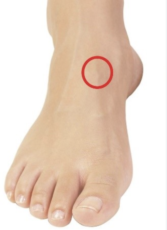
Gradual onset of vague, localised midfoot pain over the dorsal aspect of the foot.
Pain worsened by weight-bearing activities such as running or jumping.
Tenderness on palpation of the “N spot” — the dorsal, proximal area of the navicular bone.
Swelling and sometimes mild bruising at the site of injury.
Pain that improves with rest but may persist or escalate without adequate recovery.
In advanced or more serious cases, patients may experience difficulty bearing weight or an antalgic gait pattern.
Early diagnosis through clinical examination combined with imaging techniques (such as MRI or CT scans) is crucial due to the risk of progression to complete fractures or delayed healing.
This evidence-based understanding of navicular stress fractures aids clinicians and patients alike in recognising the condition promptly, facilitating targeted interventions that minimise downtime and optimise recovery outcomes.