
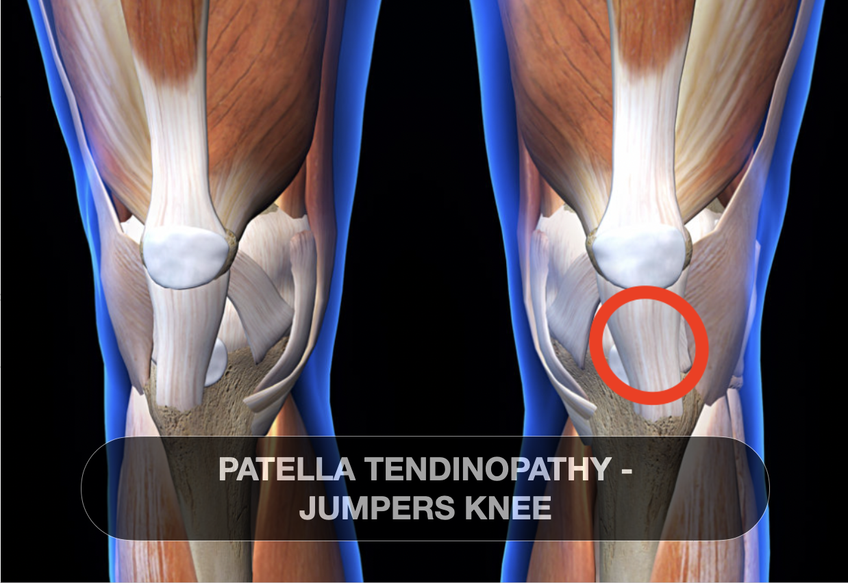
Patellar Tendinopathy - JUMPERS KNEE
🦵 Patellar Tendinopathy: Overview and Types
Definition: Patellar tendinopathy is a chronic overuse injury of the patellar tendon, characterized by pain and dysfunction at the inferior pole of the patella.
Common Types:
Jumper’s Knee: Chronic insertional injury at the inferior pole of the patella.
Sinding-Larsen-Johansson Disease: Apophysitis affecting the inferior pole of the patella, often seen in adolescents.
Osgood-Schlatter Disease: Inflammation at the tibial tuberosity, common in growing adolescents.
🧬 Pathophysiology and Classification
Histopathological Stages:
Reactive Tendinopathy: Early stage with tendon swelling and increased cellularity.
Tendon Dysrepair: Moderate stage with matrix disorganization and neovascularization.
Degenerative Tendinopathy: Late stage with collagen disorganization and tendon degeneration.
Functional Classification:
Stage I: Pain after activity, no functional impairment.
Stage II: Pain during and after activity, minor functional impairment.
Stage III: Pain during activity, significant functional impairment.
Stage IV: Chronic pain, significant functional impairment, possible tendon rupture.
🦵 Common Signs and Symptoms
Pain Localization: Anterior knee pain, localized to the inferior pole of the patella.
Aggravating Activities: Pain worsens with activities involving knee extension, such as jumping, squatting, and running.
Morning Stiffness: Stiffness and pain upon waking, improving with activity.
Tenderness: Palpable tenderness at the inferior pole of the patella.
Functional Impairment: Difficulty with activities requiring knee extension.
🏃♂️ Risk Factors
Age: Common in adolescents and young adults.
Sporting Activities: High prevalence in jumping sports like basketball, volleyball, and track and field.
Training Errors: Sudden increases in training intensity or volume.
Biomechanical Factors: Abnormal lower limb alignment and muscle imbalances.
🩺 Diagnosis and Imaging
Clinical Examination: Assessment of pain localization, tenderness, and functional limitations.
Ultrasound: Detection of neovascularization and tendon thickening.
MRI: Evaluation of tendon structure and identification of tears.
🧪 Treatment Approaches
Conservative Management:
Strengthening: ECCENTRIC :Decline squats to promote tendon remodeling. ISOMETRIC holds to decreae inhibitiona nd manage pain. Book in to see our entire strengthening plans
Load Management: Gradual increase in activity to prevent overuse.
Stretching and Flexibility Exercises: To improve muscle balance and reduce strain on the tendon.
Advanced Therapies:
Platelet-Rich Plasma (PRP) Injections: To promote healing and reduce inflammation.
Shockwave Therapy: To stimulate tendon repair and reduce pain.
Surgical Intervention: Considered in cases unresponsive to conservative treatment.