
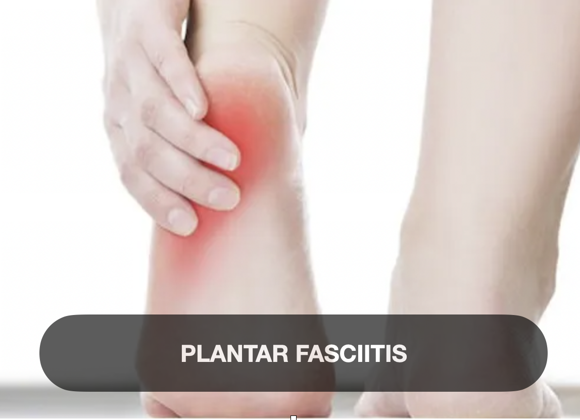
Plantar Fasciitis
Introduction
Plantar fasciitis is one of the most common causes of heel pain, affecting millions of people worldwide. Despite its prevalence, many individuals struggle with persistent discomfort that interferes with daily activities and diminishes quality of life. Understanding what plantar fasciitis is, why it occurs, and the symptoms associated with it is the essential first step toward effective treatment and healing.
What is Plantar Fasciitis?
Plantar fasciitis is an inflammation of the plantar fascia, a thick band of connective tissue that runs along the bottom of the foot from the heel bone (calcaneus) to the toes. This fascia acts like a shock absorber, supporting the arch of the foot during weight-bearing activities such as walking, running, and standing. When this tissue is overstressed or irritated, microtears develop, leading to inflammation and pain—a condition clinically known as plantar fasciitis.
Why Do People Get Plantar Fasciitis?
The development of plantar fasciitis is often multifactorial. Common causes and risk factors include:
Overuse and repetitive stress: Excessive running, walking, or standing for prolonged periods places continual strain on the plantar fascia.
Biomechanical abnormalities: Flat feet, high arches, or an abnormal gait can increase tension in the plantar fascia.
Obesity: Increased body weight places added stress on the plantar fascia.
Improper footwear: Shoes lacking adequate arch support or cushioning can exacerbate stress on the heel and foot.
Tight calf muscles or Achilles tendon: Limited flexibility in these areas can increase strain on the plantar fascia.
Age: Plantar fasciitis is most common in adults between 40 and 60 years old, as the fascia naturally loses elasticity with age.
Common Signs and Symptoms of Plantar Fasciitis
The hallmark symptom of plantar fasciitis is pain in the heel, especially near the point where the plantar fascia attaches to the heel bone. The pain is typically:
Sharp or stabbing: Often described as a sharp pain rather than a dull ache.
Worst with the first steps after rest: Many experience intense heel pain after waking up or prolonged sitting.
Improves with movement: Pain may decrease after initial activity but can worsen after prolonged standing or walking.
Localized to the bottom of the heel: While sometimes the pain may radiate along the arch, it usually centers near the heel.
Tenderness upon palpation: Pressing on the heel can elicit tenderness or discomfort.
Persistent plantar fasciitis pain can interfere with both daily activities and athletic performance. Fortunately, understanding the condition and initiating proper care can lead to significant healing and pain relief—ultimately allowing you to “heal your heel” and regain mobility.
Alright, let’s talk about the Heal Your Heel Rehab Guide for plantar heel pain. Look, if you’ve ever had that stabbing ache right under your heel that just won’t quit, you know how frustrating it is. You’re not alone. Tons of people suffer from this nagging pain — runners, desk jockeys, busy parents — you name it. But here’s the deal: this guide isn’t just some boring, cookie-cutter set of instructions. It’s built from real experience, real solutions, and real understanding of what your heel actually needs to get better.
This isn’t about quick fixes or flashy gimmicks. It’s about breaking down plantar heel pain, step-by-step, and showing you how to ease into recovery with exercises, stretches, and smart choices that actually work. It’s designed to fit your life — no fancy equipment, no confusing jargon, just clear, practical steps to help your heel heal so you can get back to doing what you love.
Most importantly, this guide gets that your story matters. Your pain story, your recovery story. It’s a living plan that grows with you — as your heel gets stronger, the guide evolves. Because your body isn’t static, and your healing shouldn’t be either.
If you’re tired of wading through a sea of websites and advice that doesn’t feel personal, this is the place where you get it straight, with clarity and confidence. You don’t have to overthink it — healing your heel is possible, and this guide is the next best step on that journey.
It’s time to stop worrying and start healing. Your heel deserves it. You deserve it.
But wait there is more
Introduction
Plantar fasciopathy—historically termed plantar fasciitis—is among the most notable causes of heel pain, affecting approximately 10 % of adults during their lifetime and contributing to 80 % of heel pain cases at any given time. This condition imposes significant physical, psychological, and socio-economic burdens, given its high prevalence among athletes, active individuals, and those with sedentary lifestyles. Contemporary research reframes its pathophysiology as a degenerative process—“fasciosis”—rather than strictly inflammatory “fasciitis” The following structured summary delves into its definition, etiology, classification, clinical features, and evolving trends in management.
1. Definition
Traditional: Chronic heel pain resulting from repetitive strain of the plantar fascia at its calcaneal origin
Modern: A non-inflammatory, degenerative condition of the plantar fascia—better termed plantar fasciosis—characterized by collagen breakdown and tissue degeneration
Clinical diagnosis: Typically based on characteristic heel pain elicited by palpation of the medial calcaneal tubercle and by dorsiflexion of the toes toward the shin; imaging (ultrasound/MRI) is used only when diagnosis is unclear or refractory to treatment
2. Cause (Etiology)
Biomechanical overload: Repetitive microtrauma from running, jumping, prolonged weight-bearing, or sudden activity increases
Intrinsic risk factors:
Elevated body mass index
Abnormal foot arch (flat or high)
Tight calf (gastrocnemius-Achilles) complex
Limited ankle dorsiflexion
Histopathology: Microtears with degenerative changes—disorganized collagen, calcifications, fibrosis
Terminology update: The term “fasciosis” is preferred over “fasciitis” to reflect degenerative rather than inflammatory pathology
3. Classification
No universally accepted staging exists, but practical categorization is based on:
Acute vs. chronic: Chronic defined by symptoms persisting beyond 3–6 months.
Recalcitrant category: Cases unresponsive to >3 months of conservative care.
Surgical vs. non-surgical candidates: Based on failure of interventions and functional limitations.
Minimally invasive options: Includes needle fasciotomy, endoscopic release, percutaneous procedures
4. Signs & Symptoms
Core complaint: Sharp, stabbing heel pain—especially with first steps after rest or at the start of activity
Typical progression: Pain subsides after 10–15 minutes of movement but may recur after prolonged standing or physical activity
Additional symptoms:
Stiffness and mild swelling
Possible pain radiating into the arch or Achilles region
Physical findings: Tenderness at medial calcaneal insertion; positive windlass stretch test
5. Current Treatment Trends
5.1 Conservative / First-line
Exercise:intrinsic foot muscle strengthening via barefoot exercises
Footwear & orthoses: Supportive shoes, over-the-counter or custom orthotics, taping, rocker shoes, night splints, and ankle-foot orthoses to reduce load
Manual interventions: Includes foot/ankle mobilizations, dry needling, eventually supported by low-level evidence
Corticosteroids: Short-term pain relief post-injection, though benefits dissipate beyond one month and may carry risks
NSAIDs and analgesia: Used symptomatically; not disease-modifying
Shockwave therapy (ESWT): Strong evidence for chronic cases unresponsive to conservative care; pain relief lasting up to one year
5.2 Emerging Therapies
Biologics:
PRP and dextrose prolotherapy show promising results, especially in recalcitrant cases; recent RCT indicated both reduce pain, with PRP slightly superior
Acupuncture: Some evidence of 4–8‑week pain relief, effects often transient; benefits similar when combined with NSAIDs
Minimally invasive procedures:
Endoscopic plantar fascia release and needle tenotomy demonstrate pain reduction over traditional care in selected refractory cases
5.3 Surgical Interventions
Open or endoscopic fasciotomy: Indicated after ≥6 months of failed conservative therapy; about 76 % success rate with minimal complications
Gastrocnemius recession: Considered in patients with calf tightness; improves dorsiflexion and outcomes .
Novel approaches: Coblation, radiofrequency ablation, and pulsed radiofrequency show early promise but need stronger evidence .
6. Summary & Clinical Implications
Natural history: 80–90 % of patients improve within 6–12 months with conservative care .
Evidence-based algorithm:
Initiate with education, load management, rehab, footwear optimization and orthoses.
If >3 months with persistent pain, escalate to ESWT, corticosteroids (short‑term), acupuncture, or biologic injections.
For cases unresponsive after 6 months, consider minimally invasive intervention or fasciotomy.
Research Gaps: Comparative efficacy, long-term safety, cost-effectiveness, and mechanistic understanding remain incomplete .
Key prognostic factors: High BMI, central pain sensitisation and psychological distress may hinder recovery; target multidimensional management .
Presentation Tips
Visuals: Include diagrams of the plantar fascia anatomy, biomechanical load charts, and a decision tree for treatment escalation.
Clinical cases: Highlight common "morning heel pain" presentations progressing to chronic symptoms.
Balance: Address both established treatments (stretching, orthoses, ESWT) and cutting-edge options (PRP, acupuncture, minimally invasive procedures) with evidence quality annotation.